Cobra 5 MP the next Generation Non Mydriatic Fundus Camera
Product description

By loading the video, you agree to YouTube's privacy policy.
Learn more
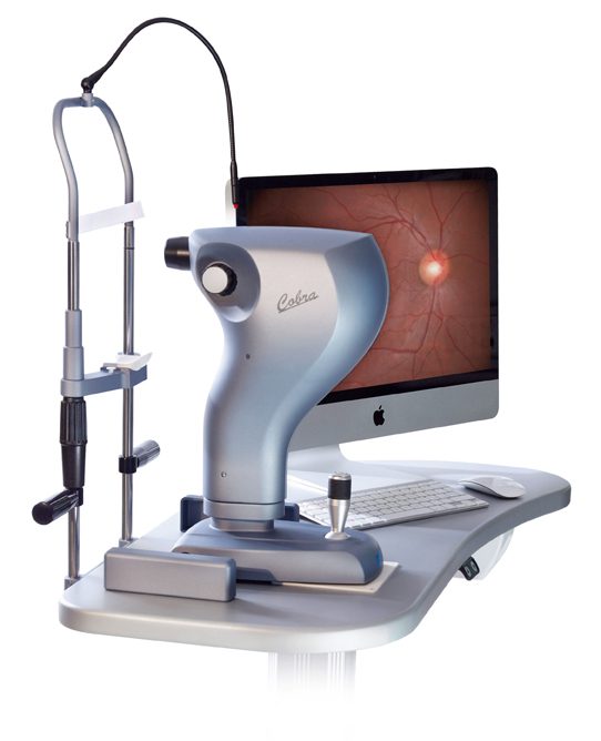
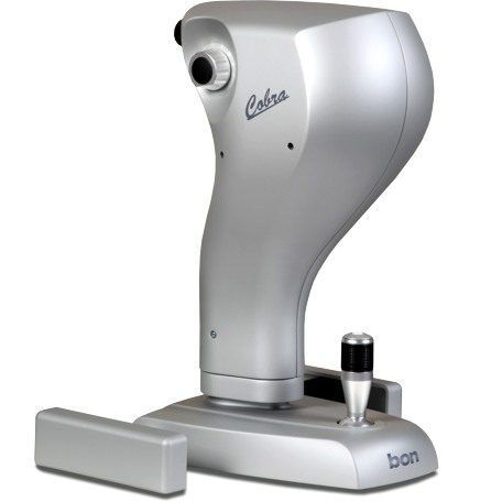
Cobra 5 MP the next generation of non-mydriatic fundus camera
The Cobra is a high resolution Fundus Camera which is ideal for fast retinal screening. The soft flash allows immediate capture of the second eye and guarantees patient comfort. With a minimum pupil size of 2.2mm excellent results can be achieved even under normal lighting conditions.
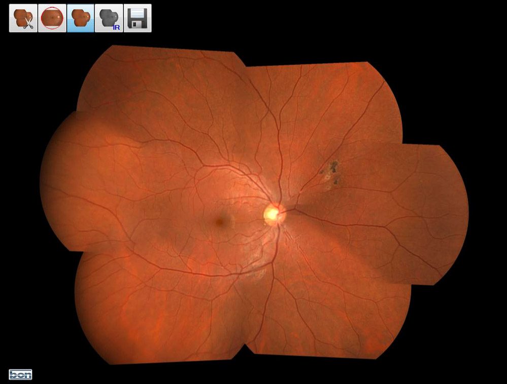
The true image has a field of view of 52 x 45 degrees which can be extended to a total of 80 x 45 degrees using the manual mosaic capture function.
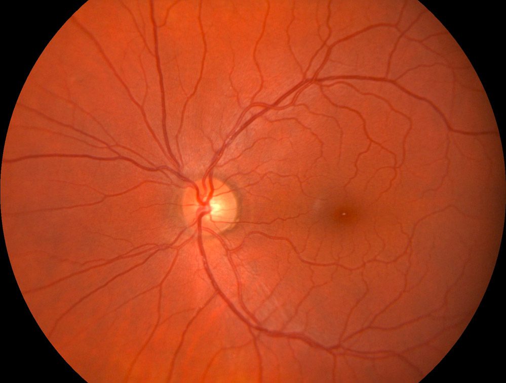
The high end CCD camera combined with the renowned CSO Optics delivers brilliant crisp images every time.
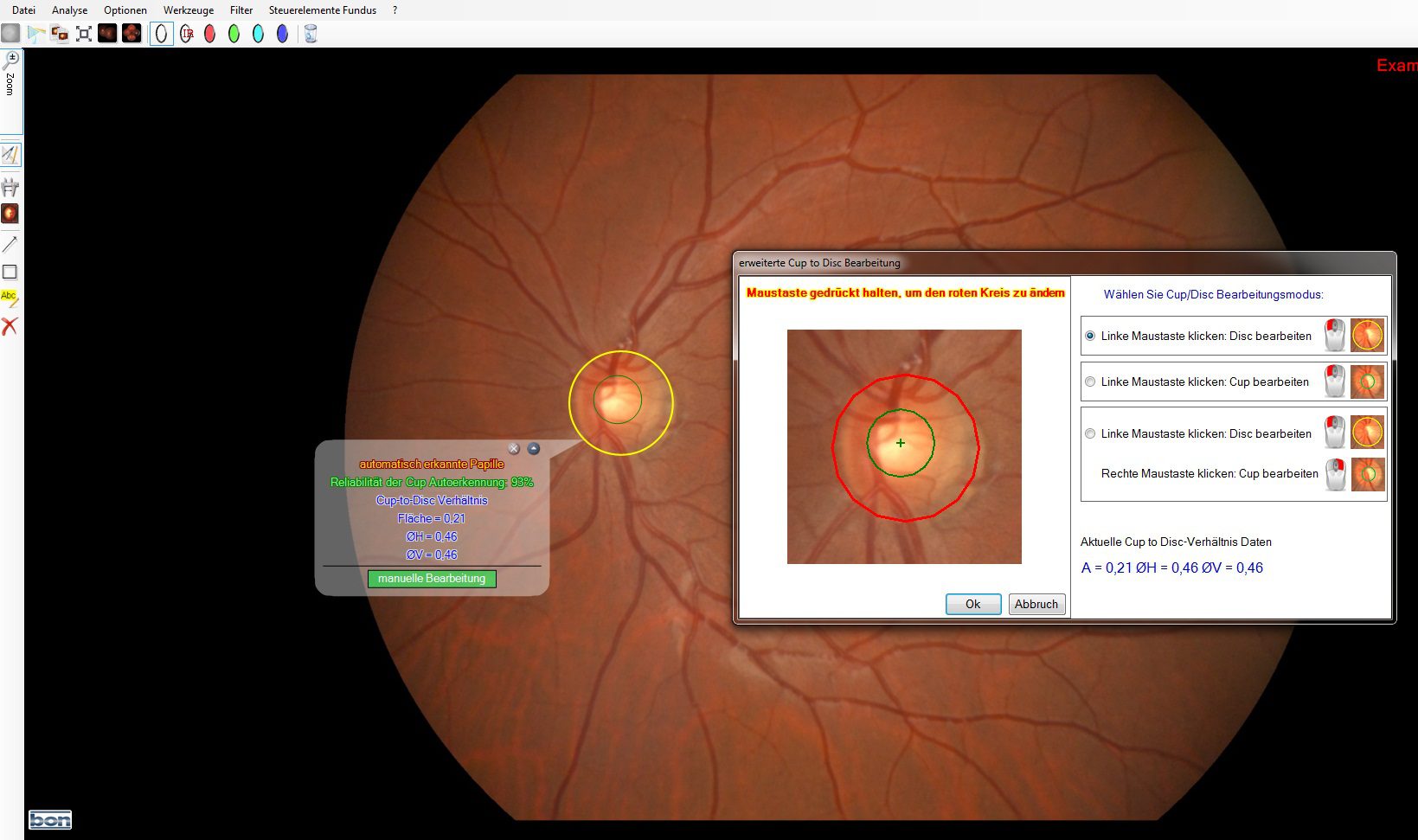
The Phoenix Software with its extensive suite of tools provides automatic cup / disc ratios, filters , full in picture editing, up to 6 picture comparison and much more.
A Retinal Vessel Analyzer is available as an option.
The compact ergonomic design fits into the smallest of testing rooms. Cobra fits easily onto the second position of your refraction unit.
Non-mydriatic
Ideal for Fundus Screening, capture great images down to 2 .2 mm pupil size even under normal ambient lighting.
High-speed image transfer to PC
Fast USB 3.0 PC connection allows quick and easy transfer of the images. The data are saved in a database through the Phoenix Software in Stand-Alone or network configuration.
DICOM connection can be achieved transferring images to a compatible server.
Anterior Segment
Thanks to its variable focus Cobra can also be used to acquire anterior segment images.
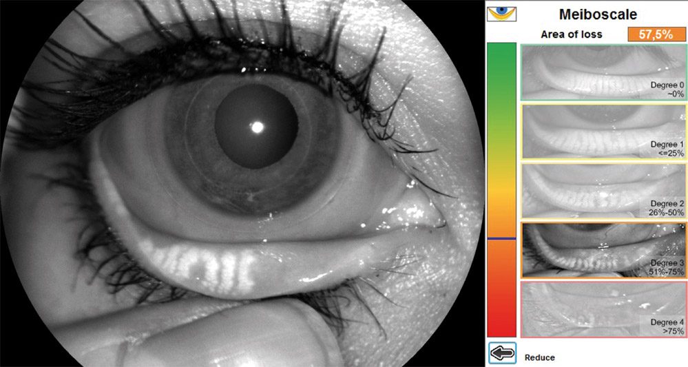
Meibography
Meibomian gland dysfunction (MGD) is the most significant cause of hyper-evaporative dry eye disease. By using infra red light the Cobra can be used to acquire images of the meibomian glands. The image can be digitally edited to highlight gland loss which can be quantified using the integral grading scale. With the Phoenix software up to 6 pictures can be compared – ideal for documentation of disease progression.
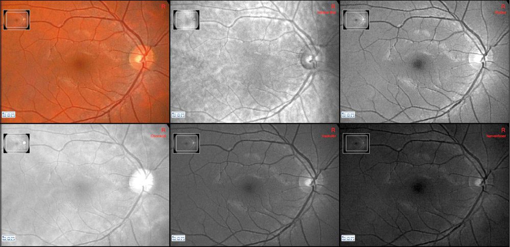
Phoenix Software imaging and archiving features
The Phoenix software incorporates a suite of editing functions including image processing, drawing, in picture measurement, zoom and text.
The manual mosaic feature allows wide field imaging for full fundus analysis.
A number of reports can be generated and saved to the patient data card.
The images can be exported in a choice of formats and Phoenix is fully compatible with DICOM.
Imaging features
- Zoom effects
- Color control
- Measurement
- C/D ratio
- Drawing (text / objects can be inserted)
- Edge enhancement
- Red-free and channel splitting
- Gamma control and filtering
- comparisons, overlay images
- automatic mosaic-mode
- Up to 6 picture comparison
- Picture overlay
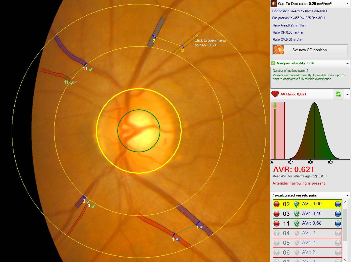
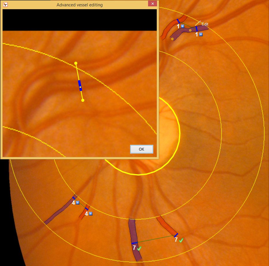
Vessel Analyser
The optional vessel analyzer allows assessment of the artery-vein ratio (A/V ratio) revealing risks of cardiovascular disease thus providing enhanced benefits for your patients.
Click on the button to load the content from Activate google recaptcha.